
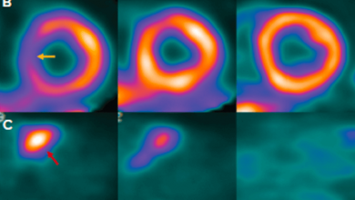


PET is a nuclear-based modality used for decades as an important clinical tool to non-invasively image blood flow, cardiac tissue metabolism and receptor expression. PET has proven to be a superior technique for molecular imaging due to its accurate attenuation correction, higher spatial and temporal resolution, higher sensitivity, quantitative abilities coupled with lower radiation risk due to the use of short-lived isotopes.
Cardiac PET has greatly improved our understanding of human coronary vasomotor function in both physiological and pathophysiological conditions. Furthermore, it has been demonstrated that the diagnostic accuracy of myocardial perfusion imaging with PET for detecting obstructive coronary artery disease surpasses that of single photon emission CT (SPECT).